Open Access Peer-Reviewed Established 1988

Pakistan Journal of Pharmaceutical Sciences

Pakistan Journal of Pharmaceutical Sciences (PJPS) is an international, peer-reviewed, open-access journal dedicated to publishing high-quality research that advances pharmaceutical, biomedical, and medicinal sciences. Published by the Faculty of Pharmacy and Pharmaceutical Sciences, University of Karachi since 1988, PJPS provides a trusted platform for researchers, academicians, clinicians, and industry professionals worldwide to disseminate innovative discoveries, foster scientific collaboration, and accelerate the translation of research into better healthcare outcomes. We welcome original research, reviews, and emerging innovations that shape the future of pharmaceutical sciences.

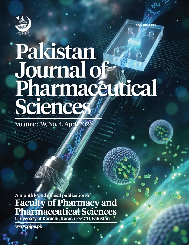
Journal Cover Vol.39
0.6
IMPACT FACTOR
Clarivate Analytics
1011-601X/3105-9686
ISSN PRINT / ONLINE
Since 1988
12400+
TOTAL CITATIONS
Google Scholar
3500+
ARTICLES PUBLISHED
Peer-Reviewed
89+
COUNTRIES REACHED
Global Readership

Latest Research Articles

Browse All Articles
original articlesPublished: 2026-11-01
Volume 39, Issue 11

Molecular mechanism of ephedrine-regulated ferroptosis in asthma via PKM2–ACSL4 lactylation

Background: Asthma is a chronic airway inflammatory disease with limited curative options. Ephedrine (EP), a natural alkaloid, has shown anti asthmatic effects, but its molecular mechanism remains incompletely understood. Ferroptosis, an iron dependent lipid peroxidation cell death pathway, aggravates airway epithelial injury in asthma. Pyruvate kinase M2 (PKM2) generates lactate, which can drive protein lactylation; however, whether EP acts by modulating PKM2-dependent lactylation of acyl-CoA synthetase long-chain family member 4 (ACSL4) to inhibit ferroptosis is unknown. Objectives: To investigated whether EP attenuates ferroptosis-driven asthmatic progression by suppressing PKM2-dependent lactylation of ACSL4. Methods: LPS-stimulated BEAS-2B cells and ovalbumin (OVA)-sensitized rats were used as in-vitro and in-vivo asthma models, respectively. Cell proliferation (CCK-8), apoptosis (TUNEL), inflammatory cytokines (ELISA) and oxidative/lipid peroxidation markers (ROS, Fe²?, MDA, LPO, SOD) were measured. PKM2 and ACSL4 expression were determined by qPCR and Western blot; ACSL4 lactylation was assessed by co-immunoprecipitation. In-vivo, asthma behavioral scores, airway resistance (RL), dynamic compliance (Cdyn), lung histology (HE) and bronchoalveolar lavage fluid cytokines were evaluated. Results: In-vitro, EP restored cell viability, reduced apoptosis and cytokine release and inhibited ferroptosis (P < 0.05). LPS upregulated PKM2; EP reversed this, whereas PKM2 overexpression abolished EP’s protection (P < 0.05). PKM2 overexpression increased ACSL4 transcription and lactylation, effects attenuated by EP. Silencing ACSL4 reversed PKM2 overexpression induced injury and ferroptosis (P < 0.05). In-vivo, EP alleviated OVA induced asthmatic symptoms and pulmonary pathological changes. Conclusion: EP mitigates asthma by down-regulating PKM2, thereby blocking ACSL4 lactylation and ferroptosis-mediated airway epithelial injury. The PKM2 ACSL4 lactylation axis represents a novel therapeutic target.

Page No:3256-3269
Lanting Zhao, Rong Xia, Shuang ZengView more
View Abstract
original articlesPublished: 2026-11-01
Volume 39, Issue 11

Xiaoshen formula delays progression of diabetic kidney disease and improves glucose and lipid metabolism: Association with the JAK/STAT pathway

Background: DKD is a leading cause of end-stage renal disease with limited therapeutic options. Objectives: This study evaluated the effects of Xiaoshen Formula (XSF) on glucose/lipid metabolism and renal injury in a DKD model of mice and preliminarily explored its mechanisms involving the JAK/STAT pathway and PPAR?. Methods: Diabetes and early-stage DKD were established in KK-Ay mice. Successfully modeled mice were randomly assigned to preventive or therapeutic administration groups; C57BL/6J mice served as blank controls. Biochemical parameters, kidney index and renal histopathology (hematoxylin-eosin staining and transmission electron microscopy) were assessed. Renal protein expression was determined by Western blot. Results: Compared with blank controls, model mice exhibited increased body weight, polydipsia, polyphagia, disrupted glucose/lipid metabolism and renal injury, confirming successful modeling. Irbesartan reduced fasting blood glucose, kidney weight, organ index and renal injury. XSF-H significantly improved glucose/lipid metabolism, reduced food/water intake and kidney weight/organ index and alleviated renal injury, with overall efficacy ranking: high-dose > therapeutic > low-dose. Western blot showed lower levels of JAK2, p-STAT-6, TGF-?1, and FN, and higher levels of PPAR? in the Irbesartan and XSF groups compared with the model group. Conclusion: This study provides experimental evidence that XSF may delay early DKD progression. XSF ameliorates fasting blood glucose, stabilizes body weight, improves glucose/lipid metabolism and attenuates renal injury and no obvious hepatorenal toxicity under limited conditions. Findings suggest that XSF delays DKD progression may be associated with modulation of the JAK/STAT signaling pathway and inhibition of renal fibrosis.

Page No:3270-3282
Han Yang, Xin Wang, Jin-ying ChenView more
View Abstract
original articlesPublished: 2026-11-01
Volume 39, Issue 11

Duo Nang Decoction enhances endometrial receptivity in PCOS rats by upregulating caveolin-1 expression

Background: Duo Nang Decoction (DND) is widely used for treating polycystic ovary syndrome (PCOS)-related infertility. However, the molecular mechanisms underlying its action remain unclear. In a previous study, downregulation of caveolin-1 (Cav-1) was shown to impair endometrial angiogenesis, a key determinant of endometrial receptivity. Objectives: This research study aimed to elucidate the influence and potential molecular mechanisms of DND on endometrial receptivity, specifically focusing on Cav-1 in PCOS rats. Methods: Sprague Dawley (SD) rats (females, 3-week-old) were randomly categorized into the DND and aspirin cohorts. After successful PCOS modeling, the rats were orally administered DND or aspirin, respectively. Then, ovulation was induced and mating was performed. The rats were euthanized on the 2nd and 5th day of conception and the endometrium was extracted for histological evaluation via hematoxylin and eosin (HE) staining. Furthermore, immunohistochemistry (IHC) staining was carried out to assess Cav-1, vascular endothelial growth factor (VEGF) and ?-catenin levels, as well as endometrial microvascular density (MVD). Moreover, reverse transcription polymerase chain reaction (RT-PCR) was used to measure the mRNA levels of VEGF and Cav-1 in the endometrium of both groups. Results: The HE and IHC staining, as well as RT-PCR analysis of endometrial tissue slices, revealed that on the 2nd and 5th day of conception, both groups indicated significantly increased endometrial thickness, MVD and elevated levels of VEGF, Cav-1 and ?-Catenin. However, the MVD in the DND group was substantially higher relative to the aspirin group. Conclusion: This study revealed that DND significantly enhanced endometrial angiogenesis and improved endometrial receptivity in PCOS rats. Mechanistically, the effects of DND on endometrial receptivity may be associated with the upregulation of Cav-1 and ?-catenin. However, the specific mechanism of action warrants further research.

Page No:3294-3301
Guozeng Wang, Wenrong Zhao, Longfei MaView more
View Abstract
original articlesPublished: 2026-11-01
Volume 39, Issue 11

Astragaloside IV attenuates high glucose-induced podocyte ferroptosis via the GSK3?/Nrf2/GPX4 pathway

Objectives: To investigate whether AS-IV alleviates high glucose (HG)-induced podocyte ferroptosis and whether this effect is associated with the GSK3?/Nrf2/GPX4 axis. Methods: Differentiated MPC-5 podocytes were exposed to HG (30 mmol/L) with or without AS-IV, the GSK3? inhibitor LY2090314, or the ferroptosis inhibitor Ferrostatin-1 (Fer-1). An osmotic control (mannitol) was included. Cell viability was quantified with the CCK-8 assay. Levels of reactive oxygen species (ROS), malondialdehyde (MDA), glutathione (GSH) and Fe²? were measured. Lipid peroxidation was detected using C11-BODIPY 581/591. Mitochondrial morphology was examined by transmission electron microscopy and protein expression was analyzed by Western blot. Results: HG exposure induced podocyte injury, characterized by decreased viability, increased oxidative stress (elevated ROS, MDA and lipid ROS), GSH depletion, iron overload and mitochondrial damage. The osmotic control did not reproduce these effects. AS-IV or Fer-1 significantly attenuated the HG-induced damage and lipid peroxidation. At the molecular level, HG downregulated Nephrin, p-GSK3? (Ser9), Nrf2 and GPX4, while upregulating total GSK3?. AS-IV treatment partially reversed these protein expression changes and produced a protective pattern similar to that of LY2090314. Conclusion: AS-IV alleviates HG-induced podocyte injury, possibly by suppressing ferroptosis and this protective effect may involve modulation of the GSK3?/Nrf2/GPX4 axis. The results offer new insights into DN pathogenesis and support AS-IV as a potential therapeutic candidate.

Page No:3302-3310
Mengya Gao, Hong Jiang, Kangya LeiView more
View Abstract
original articlesPublished: 2026-11-01
Volume 39, Issue 11

Prunasin alleviates OGD/R-induced injury in H9c2 cardiomyocytes via SRC/EGFR-AKT-FoxO-mediated mitochondrial quality control

Background: Myocardial ischemia-reperfusion injury (MIRI) is still difficult to manage clinically, mainly due to limited effective strategies that can simultaneously address oxidative stress, mitochondrial dysfunction and myocardial cell loss. Prunasin is a cyanide glycoside derived from plants of the genus Prunus. Its biological activity has been reported, but its role in ischemia-reperfusion related cardiac injury is not yet clear. Objectives: To evaluate the protective effect of prunasin in an oxygen glucose deprivation/reoxygenation (OGD/R) myocardial cell model and explore its potential mechanism. Methods: Firstly, potential targets and pathways were explored through network pharmacology and molecular docking. Then, functional validation was performed using H9c2 cells subjected to OGD/R. Evaluated cell viability, LDH release, apoptosis, ROS levels, ATP content, mitochondrial membrane potential and key signaling proteins. Results: Network pharmacology highlighted SRC, EGFR and AKT1 as core targets and molecular docking results showed stable binding between prunasin and SRC, supporting this discovery. In experiments, prunasin increased cell viability in a dose-dependent manner and reduced LDH leakage, cell apoptosis and ROS accumulation. Mitochondrial function is also protected, manifested by the recovery of ATP levels and membrane potential. Mechanistically, prunasin activates the SRC/EGFR-AKT-FxO axis, accompanied by enhanced mitochondrial biogenesis, rebalancing of fission/fusion kinetics and increased mitochondrial autophagy, collectively promoting improved mitochondrial quality control under OGD/R stress conditions. Conclusions: Prunasin regulates mitochondrial quality control through the SRC/EGFR-AKT-FxO signaling pathway, thereby reducing OGD/R-induced myocardial cell injury, indicating its potential as a candidate drug for myocardial ischemia-reperfusion intervention.

Page No:3311-3322
Jiang Li, Yaoshu Li, Chengyang XuView more
View Abstract

Browse by Topic

Research Scope & Subjects

Editorial Leadership

View Former Editors-in-Chief

Prof. Dr. Harris Shoaib

Editor-in-Chief

Faculty of Pharmacy and Pharmaceutical Sciences, University of Karachi, Karachi, Pakistan.

Specializing in Pharmacognosy and Natural Products Research, with over three decades of contribution to global pharmaceutical sciences.

2020 — PRESENT

Indexed & Abstracted in Leading Databases