★Open Access • Peer-Reviewed • Established 1988

Pakistan Journal of Pharmaceutical Sciences

Pakistan Journal of Pharmaceutical Sciences (PJPS) is an international, peer-reviewed, open-access journal dedicated to publishing high-quality research that advances pharmaceutical, biomedical, and medicinal sciences. Published by the Faculty of Pharmacy and Pharmaceutical Sciences, University of Karachi since 1988, PJPS provides a trusted platform for researchers, academicians, clinicians, and industry professionals worldwide to disseminate innovative discoveries, foster scientific collaboration, and accelerate the translation of research into better healthcare outcomes. We welcome original research, reviews, and emerging innovations that shape the future of pharmaceutical sciences.

Journal Cover 40 Issue 1
0.6
IMPACT FACTOR
Clarivate Analytics
1011-601X/3105-9686
ISSN PRINT / ONLINE
Since 1988
12500+
TOTAL CITATIONS
Google Scholar
6000+
ARTICLES PUBLISHED
Peer-Reviewed
100+
COUNTRIES REACHED
Global Readership

Latest Research Articles

Browse All Articles
original articlesPublished Date:
Volume 40, Issue 2Available Online Date: 02 Nov 2026

Predictive potential of pretreatment pepsinogen and platelet-to-lymphocyte ratios for postoperatively confirmed perineural invasion in gastric cancer patients receiving paclitaxel-based chemotherapy followed by radical surgery

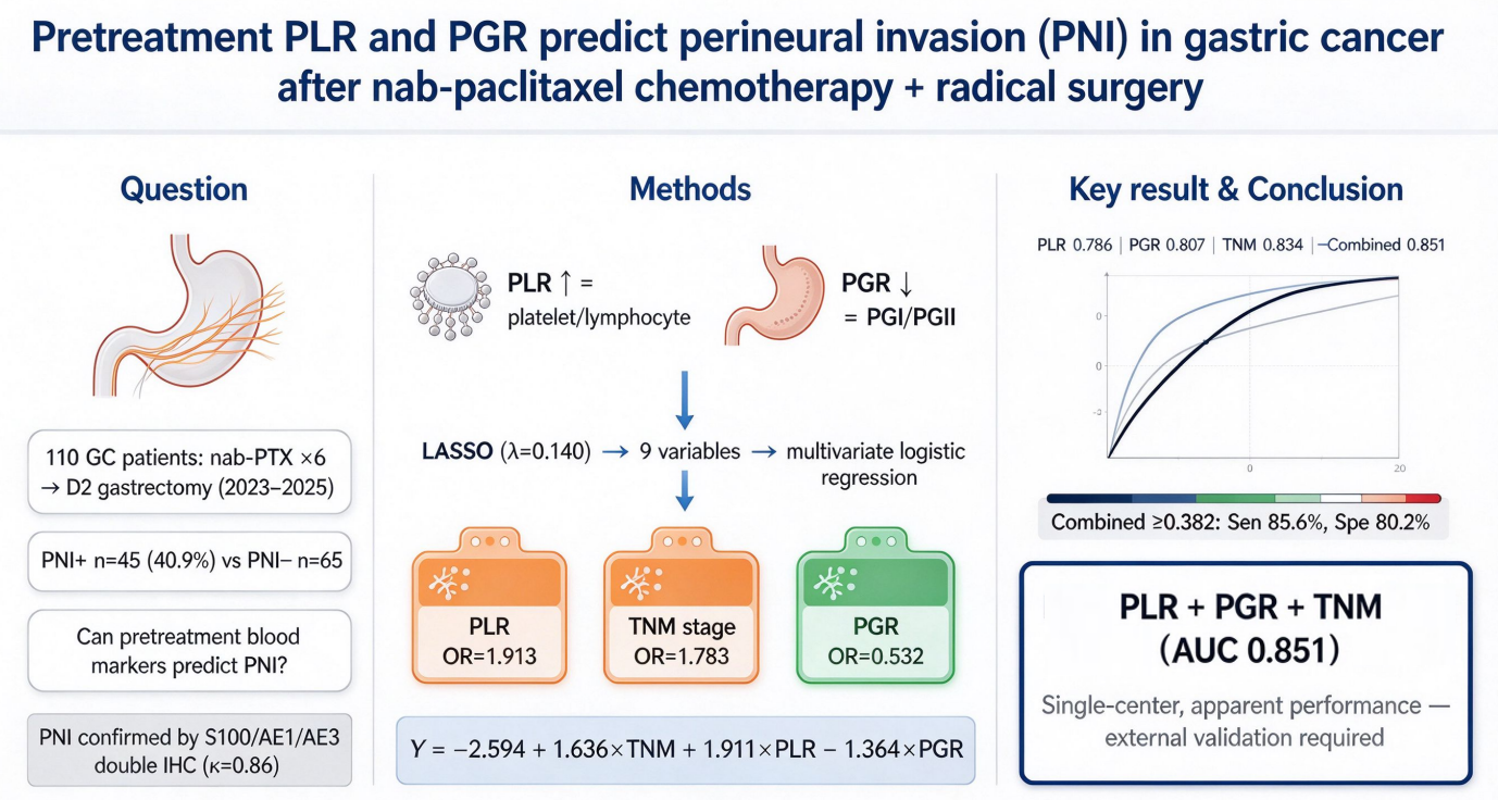
Abstract: Background: Perineural invasion (PNI) predicts poor prognosis in gastric cancer (GC) but lacks convenient preoperative biomarkers. Objectives: To investigate the predictive value of pretreatment platelet-to-lymphocyte ratio (PLR) and pepsinogen ratio (PGR) for postoperatively confirmed PNI in GC patients receiving paclitaxel-based chemotherapy followed by radical surgery. Methods: We retrospectively analyzed the medical records and clinical data of 110 patients with gastric cancer who underwent radical resection after paclitaxel-based neoadjuvant chemotherapy (from June 2023 to June 2025). Patients were divided into PNI-positive (n=45) and PNI-negative (n=65) groups. LASSO and logistic regression identified independent predictors. ROC curves, calibration, DCA and Spearman correlation were performed. Results: Of the 110 patients, 45 were PNI-positive and 65 were PNI-negative. Compared with the PNI-negative group, the PNI-positive group had significantly higher proportions of elevated CA19-9, increased tumor thickness, elevated PLR, diffuse/mixed Lauren type, Borrmann type III, T3-4 invasion, lymph node metastasis, TNM stage II/III and tumor diameter ≥5 cm, but significantly lower PGR (all P<0.05). LASSO identified nine non-zero coefficient indicators: CA19-9, tumor thickness, PLR, PGR, Lauren classification, Borrmann type III, tumor invasion, lymph node metastasis and TNM stage. Elevated PLR and advanced TNM stage were risk factors, while elevated PGR was a protective factor. ROC analysis showed apparent AUCs for PLR, TNM stage, PGR and the combined model of 0.786, 0.834, 0.807 and 0.851, respectively. PGR was negatively correlated with PNI (r = -0.749), whereas PLR (r = 0.698) and TNM stage (r = 0.751) were positively correlated. Conclusion: Pretreatment PLR and PGR show moderate predictive value for PNI in GC patients after paclitaxel chemotherapy, with slightly improved combined performance when TNM stage is added. External validation is required before clinical application.
Predictive
potential of pretreatment pepsinogen and platelet-to-lymphocyte ratios for
postoperatively confirmed perineural invasion in gastric cancer patients
receiving paclitaxel-based chemotherapy followed by radical surgery
Page No:730-743
Yongliang Li, Xi Lin, Guofeng LinView more
View Abstract
original articlesPublished Date:
Volume 40, Issue 2Available Online Date: 02 Nov 2026

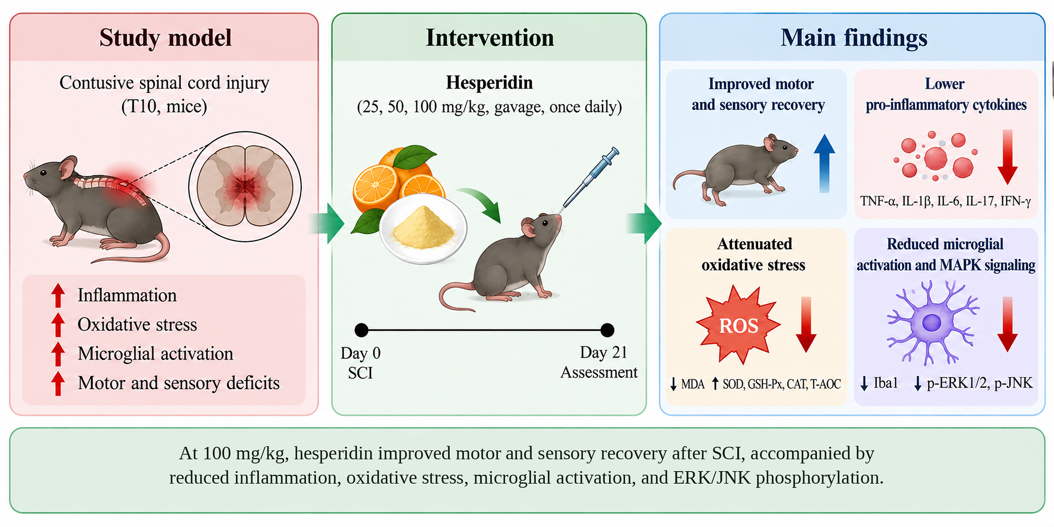
Hesperidin improves motor and sensory recovery and modulates the balance of pro- and anti-inflammatory cytokine profiles after contusive spinal cord injury in mice

Abstract: Background: Hesperidin (HES) has anti-inflammatory and antioxidant properties, but coordinated in vivo evidence linking HES to functional recovery, cytokine rebalancing, microglial activation and MAPK signaling after contusive spinal cord injury (SCI) remains limited. Objectives: To evaluate whether HES improves motor and sensory function after SCI and to characterize associated changes in the spinal inflammatory microenvironment, oxidative stress and ERK/JNK-MAPK signaling. Methods: SCI was induced by a modified Allen weight-drop contusion at T10 in 100 C57BL/6 mice randomly assigned to sham, SCI, or HES low-, medium- and high-dose groups (25, 50 and 100 mg/kg; n = 20 each). HES was administered by gavage once daily for 21 days beginning 2 h after injury. Motor and sensory outcomes were assessed by BMS scoring and mechanical and thermal tests; spinal cord cytokines, redox indices and neurorepair-related factors were measured by ELISA/biochemical assays, microglial activation by Iba1 immunohistochemistry and MAPK phosphorylation by western blotting. Results: On day 21, significant functional improvement was observed in the 100 mg/kg group (p < 0.05), whereas lower doses showed non-significant trends. In day-7 spinal cord tissue, HES reduced pro-inflammatory cytokines and increased anti-inflammatory cytokines; TNF-α decreased from 218.17 ± 28.11 to 54.39 ± 11.37 pg/mg in the high-dose group (p < 0.05). MDA levels were lower, whereas SOD, GSH-Px and CAT activities and T-AOC were higher than in the SCI group (p <0.05). HES also reduced Iba1-positive microglia and ERK1/2 and JNK phosphorylation without altering total ERK1/2 or JNK levels. Conclusion: HES was associated with improved motor and sensory recovery, a less inflammatory cytokine profile, attenuated oxidative stress and microglial activation and reduced ERK/JNK phosphorylation after contusive SCI; pathway-specific experiments remain necessary to establish causality.

Hesperidin improves motor
and sensory recovery and modulates the balance of
pro- and anti-inflammatory cytokine profiles after contusive spinal cord injury in mice
Page No:719-729
Shengli Ma, Zhiliang Yan
View Abstract
original articlesPublished Date:
Volume 40, Issue 2Available Online Date: 02 Nov 2026

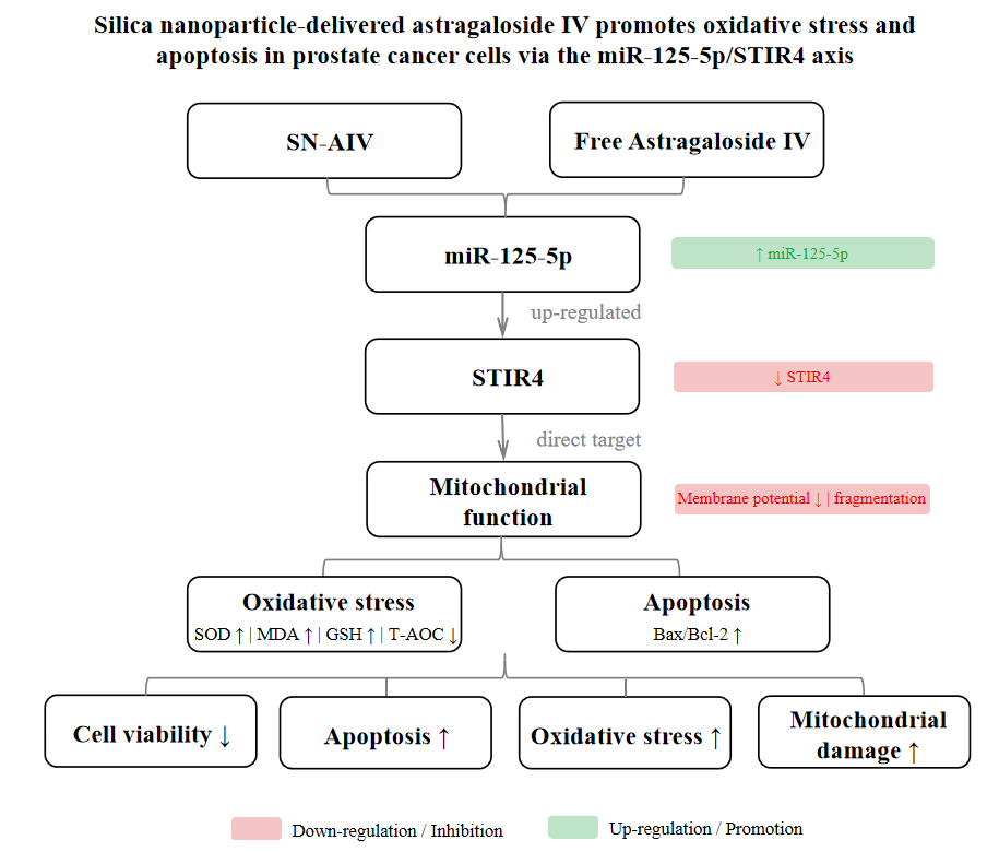
Silica nanoparticle-delivered astragaloside IV promotes oxidative stress and apoptosis in prostate cancer cells via the miR-125-5p/STIR4 axis

Abstract: Background: Prostate cancer (PCa) treatment is limited by drug resistance and toxicity. Astragaloside IV (AIV) has anti-tumor activity but poor bioavailability. Objectives: To prepare silica nanoparticle-loaded astragaloside IV (SN-AIV) and to investigate whether it induces oxidative stress and apoptosis in PC-3 cells via the miR-125-5p/STIR4 axis. Methods: SN-AIV was prepared and characterized. PC-3 cells were treated with SN-AIV, free AIV, or controls. Cell viability, apoptosis, mitochondrial function and oxidative stress markers were assessed. qRT-PCR and Western blot detected miR-125-5p and STIR4 expression. A dual-luciferase reporter assay confirmed target binding. Functional rescue experiments were performed. Results: SN-AIV exhibited uniform size (98.4 ± 6.2 nm) and sustained release. Compared with controls, SN-AIV significantly reduced cell viability, induced apoptosis, decreased mitochondrial membrane potential, increased MDA and decreased SOD and GSH levels. SN-AIV up-regulated miR-125-5p and down-regulated STIR4. STIR4 was confirmed as a direct target of miR-125-5p. miR-125-5p overexpression or STIR4 knockdown mimicked SN-AIV effects, while miR-125-5p inhibition reversed them. Conclusion: SN-AIV induces oxidative stress and apoptosis in PC-3 cells by up-regulating miR-125-5p and down-regulating STIR4, providing an experimental basis for nanoparticle-delivered AIV in PCa treatment.

Silica
nanoparticle-delivered astragaloside IV promotes oxidative stress and apoptosis
in prostate cancer cells via the miR-125-5p/STIR4 axis
Page No:705-718
Chungping Yang, Peihsin Shie, Zhenye Xu
View Abstract
reviewsPublished Date:
Volume 40, Issue 2Available Online Date: 01 Nov 2026

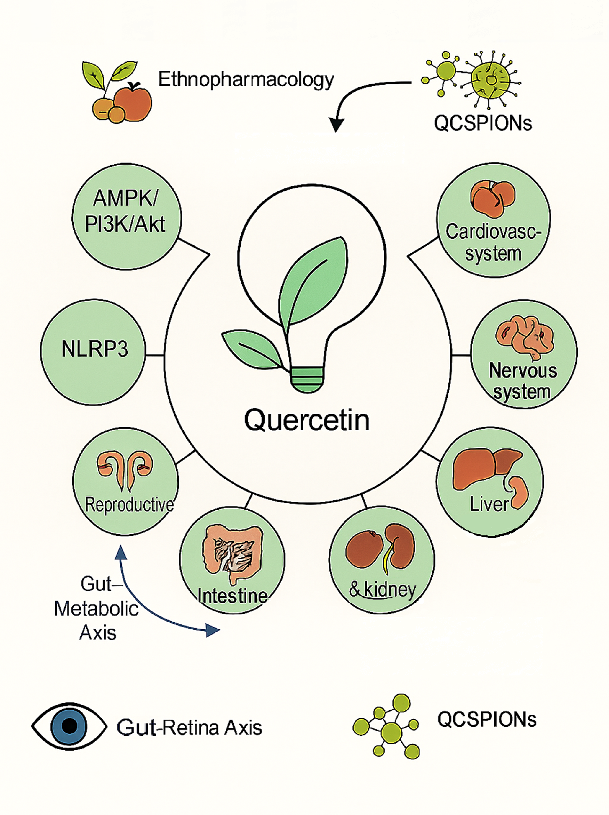
Gene–pathway–organ perspective on quercetin in diabetic complications: A narrative review of preclinical evidence

Abstract: Diabetic complications involve multiple mechanisms, including metabolic disorders, oxidative stress, inflammation, fibrosis and apoptosis. Quercetin possesses multi-target regulatory potential, but relevant preclinical evidence remains scattered across different organs and signaling pathways. This narrative review aims to synthesize rodent evidence on quercetin-related genes, signaling pathways and organ protection in diabetic complications from a gene–pathway–organ perspective. Relevant rodent studies published from inception to June 2025 were searched in PubMed, EMBASE, Web of Science, Cochrane Library and Springer Link databases. Seventy-eight studies were included after screening. Due to significant heterogeneity among studies, a narrative approach was used for evidence synthesis. Quercetin mainly regulates pathways such as AMPK, PI3K/Akt, Nrf2/HO-1, NF-κB, TGF-β/Smad3, SIRT1 and NLRP3 and is involved in metabolic, cardiovascular, hepatic, renal, nervous, intestinal, wound-healing, reproductive and retinal systems. Evidence is strong in the metabolic-cardiovascular, hepatic and renal systems, moderate in the nervous and intestinal systems and limited in other systems. Current animal evidence suggests that quercetin may act as a multi-target regulator in the intervention of diabetic complications. This "gene-pathway-organ" framework can serve as a hypothesis-generating tool to identify key molecular hubs, compare the strength of evidence across different organs and guide subsequent mechanism validation and translational research.

Gene–pathway–organ perspective on quercetin in
diabetic complications: A narrative review of preclinical evidence
Page No:694-704
Jie Zhan, Zhixin Chen, Bowen ZhangView more
View Abstract
original articlesPublished Date:
Volume 40, Issue 2Available Online Date: 29 Oct 2026

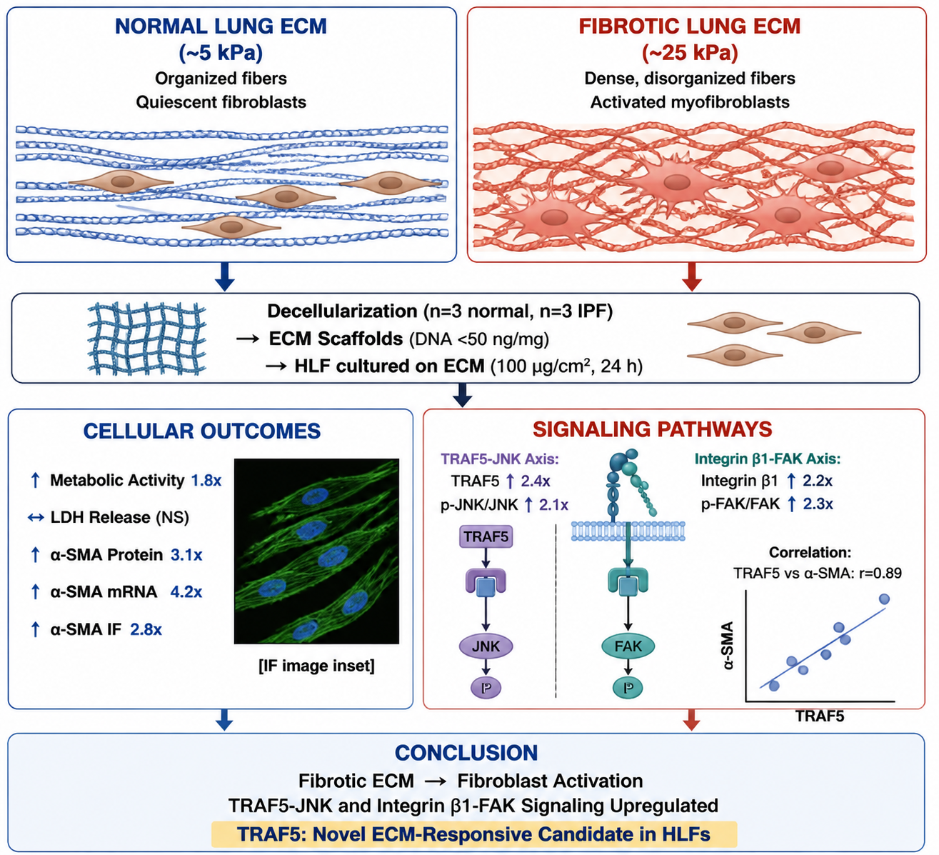
Correlational analysis of TRAF5-JNK and integrin β1-FAK signaling in human lung fibroblasts cultured on decellularized fibrotic versus normal lung extracellular matrix

Abstract: Background: Pulmonary fibrosis involves pathological remodeling of the extracellular matrix (ECM), which actively influences fibroblast behavior. The specific signaling pathways associated with fibrotic ECM-driven fibroblast activation remain incompletely defined. In particular, TNF receptor-associated factor 5 (TRAF5), a signaling adaptor traditionally studied in immune cells, has never been investigated as an ECM-responsive molecule in fibroblasts. Objectives: To investigate the effects of decellularized normal (N-ECM) and fibrotic (F-ECM) lung ECM on human lung fibroblast (HLF) metabolic activity and activation phenotype and to quantify associations with TRAF5-JNK and integrin β1-FAK signaling. Methods: Decellularized ECM scaffolds were prepared from normal (n=3) and fibrotic (IPF, n=3) human lung tissues, validated by DNA quantification (<50 ng/mg) and histology. Primary HLFs were cultured on ECM-coated plates (100 μg/cm²) for 24 hours. Metabolic activity was assessed by CCK-8 (with ECM-only controls); cytotoxicity by LDH release; protein expression by Western blot with densitometric normalization to GAPDH and total protein (p-JNK/JNK, p-FAK/FAK ratios); mRNA by qPCR; and α-SMA localization by immunofluorescence with ImageJ quantification. Pearson correlation analysis was performed to explore relationships between pathway components and activation markers. Results: F-ECM significantly increased HLF metabolic activity compared to Control (1.8-fold, 95% CI: 1.5–2.1, P<0.01) without elevating LDH release (P>0.05). ECM-only control wells showed no CCK-8 interference (OD450 < 0.05). Densitometric analysis demonstrated that F-ECM increased TRAF5 protein (2.4-fold, P<0.01), p-JNK/JNK ratio (2.1-fold, P<0.05), integrin β1 (2.2-fold, P<0.01) and p-FAK/FAK ratio (2.3-fold, P<0.01) compared to Control. The α-SMA protein (3.1-fold, P<0.01) and mRNA (4.2-fold, P<0.01) were also upregulated. TRAF5 and α-SMA protein expression showed a strong positive correlation (r=0.89, 95% CI: 0.71–0.96, P<0.001). Immunofluorescence confirmed increased α-SMA fluorescence intensity (2.8-fold, P<0.01) and spindle-shaped morphological changes in F-ECM-treated cells. Conclusion: Decellularized fibrotic lung ECM is associated with increased fibroblast metabolic activity and activation marker expression, correlating with upregulated TRAF5-JNK and integrin β1-FAK signaling. These correlational findings provide hypothesis-generating evidence for ECM-driven mechanisms in pulmonary fibrosis but require validation through pathway inhibition or gene knockdown studies to establish causality. TRAF5 is identified as a candidate ECM-responsive signaling molecule warranting further mechanistic investigation.

Correlational analysis of TRAF5-JNK and integrin
β1-FAK signaling in human lung fibroblasts cultured on decellularized fibrotic
versus normal lung extracellular matrix
Page No:682-693
YuMei Liu, Jing Shao, Meng ZhaoView more
View Abstract

Browse by Topic

Research Scope & Subjects

Editorial Leadership

View Former Editors-in-Chief

Prof. Dr. Harris Shoaib

Editor-in-Chief

Faculty of Pharmacy and Pharmaceutical Sciences, University of Karachi, Karachi, Pakistan.

Specializing in Pharmacognosy and Natural Products Research, with over three decades of contribution to global pharmaceutical sciences.

2020 — PRESENT

Indexed & Abstracted in Leading Databases