★Open Access • Peer-Reviewed • Established 1988

Pakistan Journal of Pharmaceutical Sciences

Pakistan Journal of Pharmaceutical Sciences (PJPS) is an international, peer-reviewed, open-access journal dedicated to publishing high-quality research that advances pharmaceutical, biomedical, and medicinal sciences. Published by the Faculty of Pharmacy and Pharmaceutical Sciences, University of Karachi since 1988, PJPS provides a trusted platform for researchers, academicians, clinicians, and industry professionals worldwide to disseminate innovative discoveries, foster scientific collaboration, and accelerate the translation of research into better healthcare outcomes. We welcome original research, reviews, and emerging innovations that shape the future of pharmaceutical sciences.

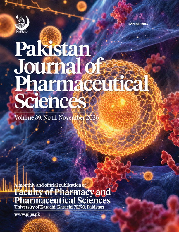
Journal Cover 39 Issue 11
0.6
IMPACT FACTOR
Clarivate Analytics
1011-601X/3105-9686
ISSN PRINT / ONLINE
Since 1988
12500+
TOTAL CITATIONS
Google Scholar
6000+
ARTICLES PUBLISHED
Peer-Reviewed
100+
COUNTRIES REACHED
Global Readership

Latest Research Articles

Browse All Articles
original articlesPublished: 01 Jan 2027
Volume 40, Issue 1

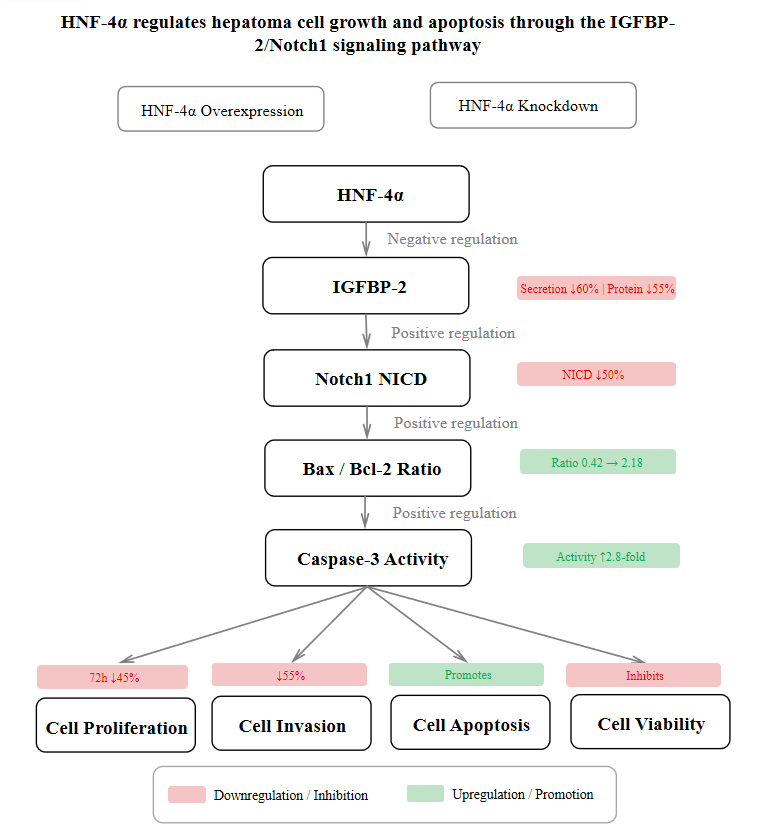
HNF-4α regulates hepatoma cell growth and apoptosis through the IGFBP-2/Notch1 signaling pathway

Abstract: Background: Hepatocyte nuclear factor 4α (HNF-4α) is a liver-enriched transcription factor that is downregulated in hepatocellular carcinoma (HCC). However, the molecular mechanisms by which it regulates the malignant phenotype of HCC cells remain unclear. Objectives: To investigate whether HNF-4α affects HCC cell proliferation, invasion and apoptosis by regulating the IGFBP-2/Notch1 signaling pathway. Methods: HNF-4α was overexpressed and silenced in HepG2 cells using a pcDNA3.1-HNF-4α plasmid and HNF-4α-specific siRNA, respectively. RT-qPCR and Western blotting verified transfection efficiency. Cell proliferation and invasion were assessed by MTT and Transwell assays, respectively. Apoptosis was evaluated by Caspase 3 activity assay. IGFBP-2 secretion was measured by ELISA. Protein expression of Bcl-2, Bax, IGFBP-2 and Notch1 was detected by Western blotting. Results: HNF-4α overexpression significantly inhibited HepG2 cell proliferation (by approximately 35% at 48 h and 45% at 72 h) and invasion (by approximately 55%), while markedly increasing Caspase 3 activity (approximately 2.8-fold) and the Bax/Bcl-2 ratio (from 0.42 to 2.18). Moreover, HNF-4α overexpression significantly suppressed IGFBP-2 secretion (by approximately 60%) and downregulated IGFBP-2 (by approximately 55%) and Notch1 NICD (by approximately 50%) protein expression (all P < 0.001). Conversely, HNF-4α knockdown produced opposite effects, significantly promoting proliferation (by approximately 40% at 48 h and 55% at 72 h) and invasion (by approximately 70%), decreasing Caspase 3 activity (by approximately 55%) and the Bax/Bcl-2 ratio (from 0.45 to 0.19) and upregulating IGFBP-2 (approximately 2.2-fold) and Notch1 NICD (approximately 1.8-fold) expression (all P < 0.01). Conclusion: HNF-4α inhibits HCC cell proliferation and invasion and promotes apoptosis through negative regulation of the IGFBP-2/Notch1 signaling pathway, providing new experimental evidence for the tumor-suppressive mechanism of HNF-4α in HCC.
HNF-4α regulates hepatoma
cell growth and apoptosis through the IGFBP-2/Notch1 signaling pathway
Page No:62-75
Changxi Liao, Xiaojie Jiang, Jianfeng ZhaoView more
View Abstract
original articlesPublished: 01 Jan 2027
Volume 40, Issue 1

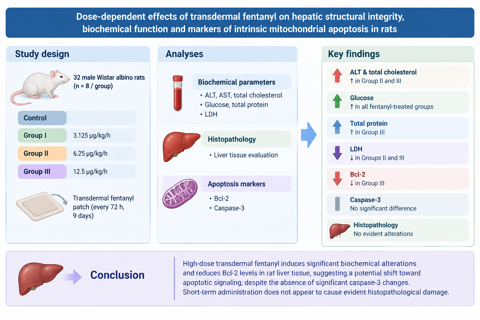
Dose-dependent effects of transdermal fentanyl on hepatic structural integrity, biochemical function and markers of intrinsic mitochondrial apoptosis in rats

Abstract: Background: Transdermal fentanyl is widely used for pain management; however, whether high systemic doses exert direct hepatotoxic effects or trigger intrinsic liver apoptotic pathways independently of systemic hypoxia remains unclear. Objectives: This study aimed to investigate the dose-dependent effects of transdermal fentanyl on hepatic structural integrity, biochemical function and markers of intrinsic mitochondrial apoptosis (Bcl-2 and caspase-3) in rats. Methods: Thirty-two Wistar albino rats were randomly assigned to four groups (n = 8): a control group and three experimental groups treated with transdermal fentanyl at doses of 3.125 µg/kg/h (Group I), 6.25 µg/kg/h (Group II) and 12.5 µg/kg/h (Group III). Patches were applied to the abdominal region and replaced every 72 hours for nine days. At the end of the experiment, liver tissues were collected for biochemical and histopathological and molecular analyses of apoptotic markers. Apoptotic changes were assessed by measuring caspase-3 and Bcl-2 levels. Results: Serum alanine aminotransferase (ALT) and total cholesterol levels were significantly higher in Group II and III than in the control group (p<0.05). Glucose levels were significantly increased in all fentanyl-treated groups (p<0.05). Total protein levels were significantly elevated in Group III compared to the control group (p<0.05). Lactate dehydrogenase (LDH) levels were significantly lower in Groups II and III (p<0.05). The anti-apoptotic marker Bcl-2 was significantly decreased in Group III compared with the other groups (p<0.05), whereas no statistically significant differences were observed in caspase-3 levels among any of the groups. No evident histopathological alterations were detected in liver tissues. Conclusion: High-dose transdermal fentanyl induces significant biochemical alterations and reduces Bcl-2 levels in rat liver tissue, suggesting a potential shift toward apoptotic signaling despite the absence of significant caspase-3 changes. However, short-term administration does not appear to cause evident histopathological damage.

Dose-dependent effects of transdermal fentanyl on
hepatic structural integrity, biochemical function and markers of intrinsic
mitochondrial apoptosis in rats
Page No:55-61
Zeynep Ozdemir, Nurettin Kurt, Omer Faruk KelesView more
View Abstract
original articlesPublished: 01 Jan 2027
Volume 40, Issue 1

Amlodipine besylate and valsartan combined mucoadhesive buccal patch formulations: In-vitro release and ex-vivo permeation studies

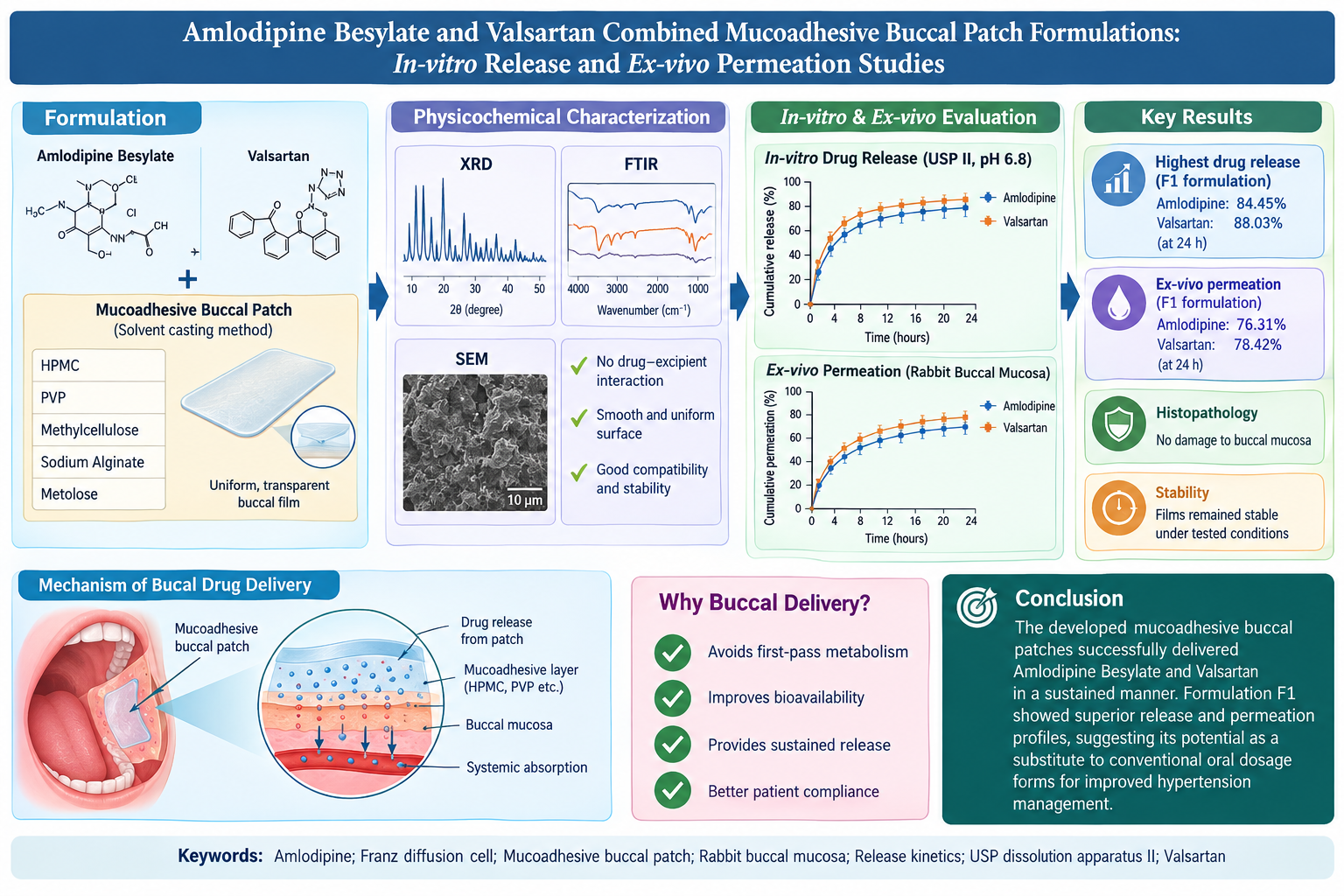
Abstract: Background: Oral fixed-dose combinations of Amlodipine Besylate and Valsartan are widely prescribed for hypertension but are associated with first-pass metabolism and variable bioavailability. Buccal drug delivery provides a favorable substitute to improve systemic availability and provide sustained release. Objectives: This study aimed to develop and assess mucoadhesive buccal patches containing Amlodipine Besylate and Valsartan to enhance bioavailability and achieve controlled drug release over 24 hours. Methods: Buccal patches were developed via the solvent-casting method using Hydroxypropyl Methylcellulose, Polyvinylpyrrolidone, Methylcellulose, Sodium Alginate and Metolose as backing membrane polymers. All the prepared films were evaluated for physicochemical parameters by means of X-ray Diffraction (XRD), Fourier transform infrared spectroscopy (FTIR), Scanning electron microscopy (SEM) and in vitro and ex vivo drug release. In-vitro drug release was studied with USP Apparatus II (paddle method) in 500 ml 6.8 pH phosphate buffer at 37 ± 0.5°C and 50 rpm for 24 hours. Ex vivo permeation studies were performed using a Franz diffusion cell fitted with rabbit buccal mucosa under similar conditions. Results: The formulations generally exhibited acceptable physicochemical characteristics within the evaluated parameters. The developed films appeared transparent with a uniform distribution of components. FTIR analysis did not indicate any detectable incompatibility or significant interaction between the drugs and excipients under the study conditions. Among all formulations, F1 contained the highest drug release and permeation. After 24 hours, cumulative release reached 84.45% for amlodipine and 88.03% for valsartan. Ex-vivo permeation was 76.31% and 78.42% for amlodipine and valsartan, respectively. Furthermore, buccal films' histopathological and stability studies were found to be harmless and functioned as an efficient dosage system. Conclusion: The developed mucoadhesive buccal patches successfully delivered both drugs in a sustained manner. Formulation F1 showed superior release and permeation profiles, suggesting its potential as substitute to conventional oral dosage forms for improved hypertension management.

Amlodipine besylate and valsartan combined
mucoadhesive buccal patch formulations: In-vitro release
and ex-vivo permeation studies
Page No:40-54
Nosheen Anwar, Rahman Gul, Syed Umer JanView more
View Abstract
original articlesPublished: 01 Jan 2027
Volume 40, Issue 1

Chemical fingerprinting and simultaneous determination of main compounds combined with multivariate analysis of the Syringa pubescens Turcz. leaves from different origins

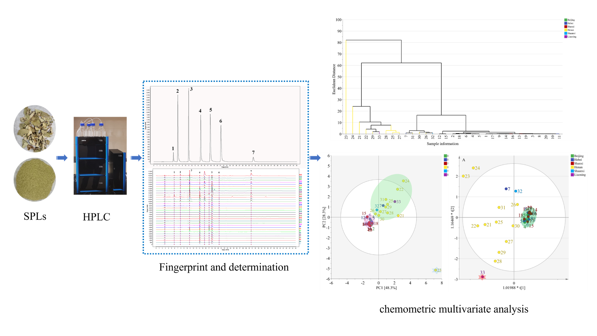
Abstract: Background: Syringa pubescens Turcz. is a folk medicinal plant in Henan Province, China and its leaves have been used for medicinal purposes. However, the bioactive compound content varies with geographical origin and quality control methods remain limited. Objectives: This aim is to develop a reliable HPLC method for the simultaneous determination of seven bioactive compounds in S. pubescens leaves from different origins and to evaluate sample differences using chemometric analysis. Methods: Thirty-three batches of leaves from six main regions in China were analyzed using HPLC. Hierarchical cluster analysis (HCA), principal component analysis (PCA) and orthogonal partial least squares discriminant analysis (OPLS-DA) were employed to the quantitative data. Results: All seven components showed good linearity (R² > 0.999). Recovery ranged from 98.23% to 100.12% and RSD values for precision and stability were below 1.84%. The contents of echinacoside, verbascoside and oleuropein were higher than those of other ingredients. HCA and PCA classified the 33 batches into three groups according to their geographical origin. OPLS-DA further differentiated the samples, with oleuropein, echinacoside, verbascoside and syringin identified as major discriminating markers. Conclusion: These findings could support the resource development and utilization and provide a reference for the quality control of SPLs.

Chemical fingerprinting and simultaneous determination of main
compounds combined with multivariate analysis of the Syringa
pubescens Turcz. leaves from different origins
Page No:32-39
Xinyi Guan, Suya Zhang, Yitong ShiView more
View Abstract
original articlesPublished: 01 Jan 2027
Volume 40, Issue 1

Effect of new zaogong erteng decoction on embryo implantation in mice following treatment of endometritis

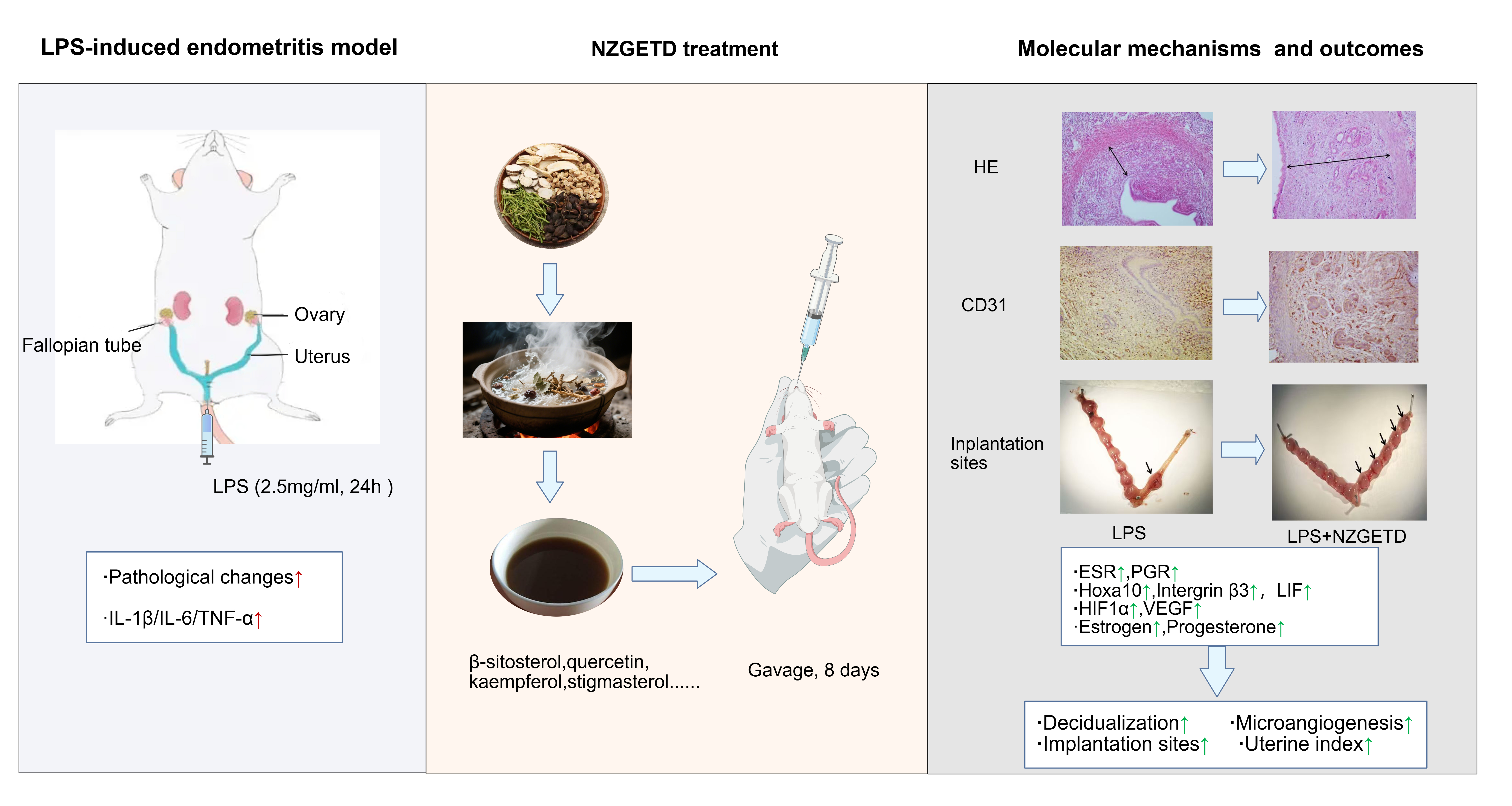
Abstract: Background: New zaogong erteng decoction (NZGETD) is a compound Chinese medicine modified from the Zaogong Erteng Decoction. Previous clinical observations suggested its potential efficacy against endometritis, an inflammatory condition that adversely impacts endometrial receptivity and embryo implantation. Objectives: In this study, the role and mechanism of NZGETD in improving embryo implantation were investigated in a mouse model of lipopolysaccharide-induced endometritis. Methods: Female mice were treated with 2.5 mg/mL lipopolysaccharide in the endometrium for 24 h to induce endometritis and were subsequently gavaged with NZGETD for 8 days. Pathological changes, inflammatory cytokine levels, uterine index, endometrial decidualization, microangiogenesis and hormone levels were assessed. Network pharmacology analysis was employed to identify potential active components and targets. The expression of key proteins related to endometrial receptivity and implantation was confirmed by western blotting. Results: NZGETD attenuated lipopolysaccharide-induced pathological changes and downregulated uterine levels of IL-1β, IL-6 and TNF-α, indicating its anti-inflammatory effect in this endometritis model. NZGETD significantly increased the uterine index, promoted endometrial decidualization and microangiogenesis (upregulated CD31 on gestational day 4.5). Additionally, NZGETD significantly elevated uterine levels of estrogen and progesterone and increased the number of implantation sites on gestational day 8.5. Network pharmacology analysis screened beta-sitosterol, quercetin, kaempferol and stigmasterol as the common active components of NZGETD. Western blotting confirmed that NZGETD up-regulated ESR, PGR, Hoxa10, integrin β3, LIF, HIF1α and VEGF during embryo implantation. Conclusion: In a mouse model of lipopolysaccharide-induced endometritis, NZGETD promoted decidualization and microangiogenesis of the endometrium, enhanced endometrial receptivity and facilitated embryo implantation.

Effect of new zaogong
erteng decoction on embryo implantation in mice following treatment of
endometritis
Page No:16-31
Shuang Li, Huiling Liu, Nan KangView more
View Abstract

Browse by Topic

Research Scope & Subjects

Editorial Leadership

View Former Editors-in-Chief

Prof. Dr. Harris Shoaib

Editor-in-Chief

Faculty of Pharmacy and Pharmaceutical Sciences, University of Karachi, Karachi, Pakistan.

Specializing in Pharmacognosy and Natural Products Research, with over three decades of contribution to global pharmaceutical sciences.

2020 — PRESENT

Indexed & Abstracted in Leading Databases